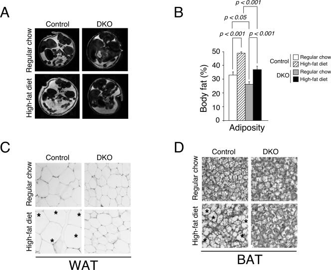
FIG. 3.
Nfatc2−/− Nfatc4−/− mice exhibit reduced adiposity. Cross-sectional image of 12-week-old Nfatc2−/− Nfatc4−/− mice (DKO) and control Nfatc2−/+ Nfatc4−/+ mice by magnetic resonance imaging (A). The measured in vivo adiposity was also shown (B). Pathohistological analysis of white (WAT) (C) or brown (BAT) (D) adipose tissues of DKO and control mice was also shown. Effect of high-fat-diet-elicited obesity was indicated. Asterisks illustrate enlarged adipocytes.

