FIG. 3.
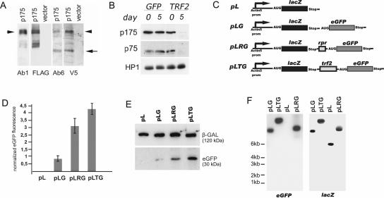
The origin of TRF2 polypeptides. (A) Western blot analysis of protein extracts from Schneider-2 cells transfected with a plasmid encoding p175 tagged with three N-terminal FLAG epitopes and a C-terminal V5 epitope (p175) and from control cells transfected with an empty vector. The antibodies used for visualization are indicated below. The protein corresponding to full-length p175 is indicated by an arrowhead, and the C-terminal polypeptide (75 kDa) is indicated by an arrow. In addition to the full-length p175, the anti-FLAG antibodies and Ab1 revealed protein species of lower molecular weight that may be the result of nonspecific proteolysis. (B) The TRF2 N-terminal and C-terminal antibodies reveal depletion of both p175 and p75 after RNAi of the trf2 gene. S2 cells were transfected with dsRNA corresponding to the N-terminal part of p175 or GFP (control) as indicated above the blots. Five days after transfection, total extracts from equivalent numbers of cells were analyzed by Western blotting with Ab1 and Ab6 and antibodies against HP1 (control). (C to F) Test of the trf2 fragment for the ability to sustain the internal translation initiation. (C) Schematic representation of constructs: pL, monocistronic reporter vector pAc containing lacZ gene; pLG, dicistronic construct containing lacZ and eGFP as the upstream and downstream cistrons, respectively; pLRG, dicistronic construct containing the rpr IRES between the lacZ and eGFP cistrons; pLTG, dicistronic construct containing the trf2 2.3-kb fragment between the cistrons. (D) Diagram illustrating eGFP fluorescence intensity in transfected cell lines normalized to the corresponding β-Gal activity. (E) Western blot analysis of protein extracts from transfected cell lines with antibodies against β-GAL and eGFP. (F) Northern blot analysis of dicistronic mRNA expression. RNA was isolated from S2 cells transfected with pL, pLG, pLRG, and pLTG constructs. Probes specific for eGFP and lacZ coding regions were sequentially used for hybridization. The positions of the molecular markers are indicated.

