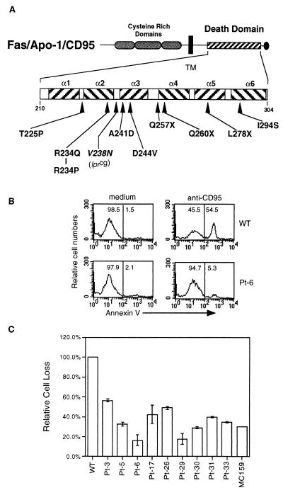
Figure 1.
CD95 “death-domain” amino acid changes in ALPS patients. (A) Diagram of the CD95 protein. The extracellular cysteine-rich domains (elongated dark ovals), the transmembrane domain (black box), the death domain (hatched box), and a putative negative regulatory domain (oval) are shown, with the positions of amino acid changes caused by the nine mutations described in this study based on the human CD95 sequence (GenBank accession nos. M67454 and X63717) (11–15, 23, 24). Also shown is the position equivalent to the mouse lprcg mutation. (B) Transient transfection assay for dominant interference in Jurkat T cells by using annexin binding as a marker of apoptosis. Flow cytometry profiles of Jurkat T-cells transfected with either WT (Upper) or a representative mutant (Pt. 6) (Lower) CD95 along with the murine class I DNA (H-2 Ld-pSRa) transfection control marker. Twelve hours after transfection, the cells were treated with either medium alone or with 30 ng/ml CH11 for 10 hr. Cells gated for H-2 Ld expression were stained with annexin V-FITC (x axis). (C) Quantitation of dominant interference with apoptosis by CD95 alleles from the patients in this study. Percent apoptosis was measured with flow cytometry as in B, and apoptotic cell loss from anti-CD95 treatment was calculated as described in Materials and Methods. Data are representative of three independent experiments and standard deviations are shown except in cases where they are too small to be depicted.

