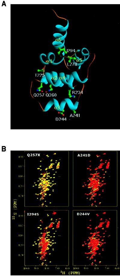
Figure 2.
(A) Ribbon diagram of the death-domain structure illustrating the location of the amino acid changes that were analyzed (9). (B) NMR comparison of the WT and ALPS CD95 death domains. HSQC spectra of bacterially synthesized death domains from four representative ALPS patients as indicated; full data for all nine patients are given in Table 1. The 15N/1H amide chemical shifts from the WT protein are shown in yellow, and those of the mutant CD95 proteins are overlaid in red. Noncoincidence of the spots indicates structural deviations from the WT.

