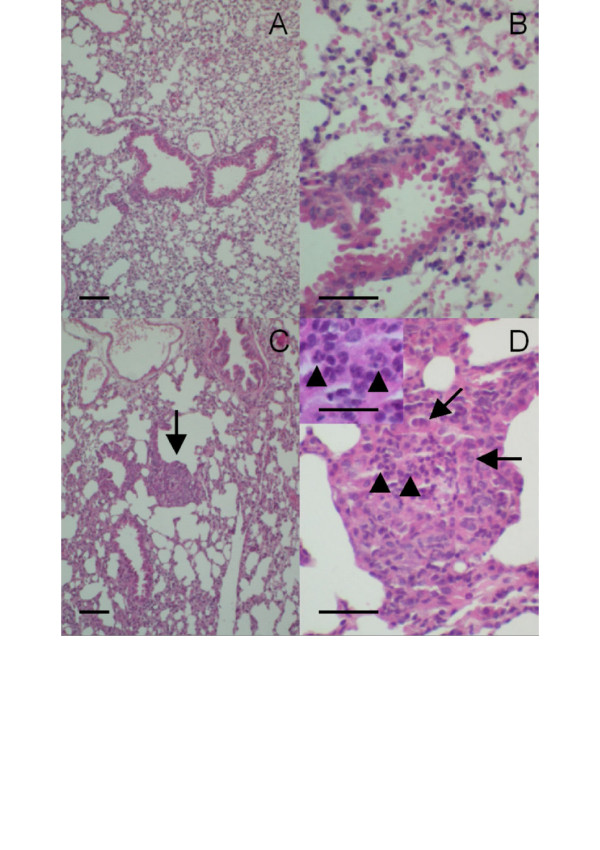
Figure 3.
Hematoxylin and eosin-stained lung sections obtained from: (A-B) a naive wild-type mouse showing no pathological abnormalities; (C-D) a naive CF mouse showing focal areas of bronchopneumonia with neutrophil infiltration (arrow in C; focus enlarged at D). Arrowheads identify neutrophils in the bronchial lumen at D and in the inset; bronchial epithelial cells are identified by arrows. Calibration bars correspond to 50 μm in panels A-D and to 25 μm in the inset.

