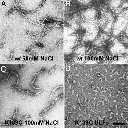
Fig. 2.
Electron micrographs of negatively stained vimentin samples. (A and B) WT assembled at ≈5 mg/ml in 5 mM Tris·HCl buffer (pH 8.4) in the presence of 50 and 100 mM NaCl, respectively. (C) K139C mutant at ≈5 mg/ml in 5 mM Tris·HCl (pH 8.4) with 100 mM NaCl. (D) K139C mutant assembled for 2 s under standard conditions (0.1 mg/ml protein, 25 mM Tris·HCl, pH 7.5, and 50 mM NaCl). (Scale bar: 100 nm.)

