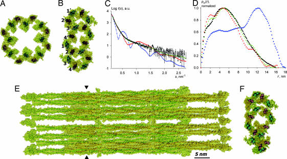
Fig. 4.
Vimentin ULF. (A) Cross-section of the initial model featuring a 4-fold symmetrical arrangement of octamers. (B) Refined model with a flattened cross-section. (C) Calculated scattering from the initial symmetrical model (blue diamonds), flattened model (red squares), and the final randomized model (green triangles) overlaid with the experimental data for the K139C mutant vimentin variant at 4 mg/ml in 5 mM Tris·HCl buffer (pH 8.4) with 75 mM NaCl (black circles). (D) Corresponding pcr(r) functions. (E) Side view of the final randomized model. (F) Cross-section of the position indicated by triangles in E in the final randomized model.

