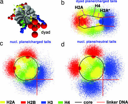
Fig. 2.
Reference frame of a nucleosome core (a) and positional distribution of fully charged histone tails along its dyad (b) and nucleosomal planes (c) and of neutralized tails along a nucleosome plane (d), all at 0.2 M salt. Red arrows in b indicate the mean position/orientation of H4 and H2A tails. H2A*, C termini of H2A histones.

