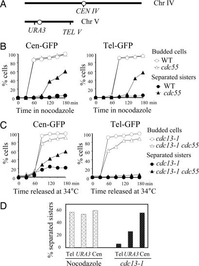
Fig. 4.
Sister chromatid separation in Δcdc55 mutants at different loci along the chromosomes after spindle or DNA damage. (A) Diagram showing the location of TetO arrays in chromosome IV and V. (B) Sister chromatid separation in Δcdc55 mutants at centromere and telomere in the presence of nocodazole. WT and Δcdc55 cells carrying tetO arrays at centromere or telomere were arrested in G1 phase and then released into nocodazole (20 μg/ml) medium at 30°C. (Left) Kinetics of Cen-GFP separation and the budding index. (Right) Kinetics of Tel-GFP separation and the budding index. (C) Centromeric and telomeric sister chromatid separation in cdc13–1-arrested Δcdc55 mutants. cdc13–1 and cdc13–1 Δcdc55 mutant cells carrying tetO arrays at centromere, as well as those carrying the tetO arrays at telomere, were first arrested in G1 phase at 25°C and then released at 34°C. The budding index and the percentage of cells with separated GFP dots are shown. (D) Comparison of sister chromatid separation rates at telomere, the URA3 locus, and centromere in Δcdc55 cells arrested by nocodazole and DNA damage. Δcdc55 (Left) and cdc13–1 Δcdc55 (Right) carrying tetO arrays at different loci were treated as described in A and B. Shown are percentages of GFP-dot separation in cells collected 180 min after release.

