Fig. 1.
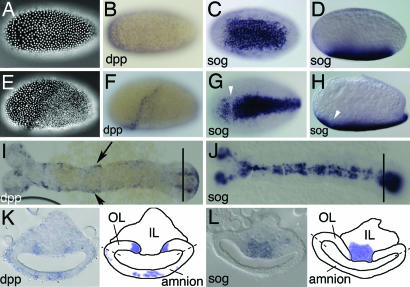
Expression of Tc-dpp compared with expression of Tc-sog. (B–D% and F–L%) In situ hybridizations with Tc-dpp (B, F, I, and K) and Tc-sog (C, D, G, H, J, and L). (A–D%) Uniform blastoderm stages. (E–H%) Differentiated blastoderm stages. (I and J) Extending germ-band embryos. (K and L) Cross-sections from the growth zone, taken at the position of the lines in I and J, respectively. Schematic drawings are shown at Right. Dashed lines indicate the future border between amnion and embryo proper (A) DAPI counterstaining of the embryo shown in B. The nuclei have a uniform distribution. (B) Lateral view. Tc-dpp is ubiquitously expressed, with stronger expression at the anterior pole. (C) Ventral view. Tc-sog is expressed in a broad ventral domain. (D) Lateral view of the embryo shown in C. (E) DAPI counterstaining of the embryo shown in F. The serosa can be recognized by big, widely spaced nuclei; the germ rudiment by smaller, dense nuclei. (F) Lateral view. Tc-dpp is expressed in a stripe along the germ rudiment/serosa border. (G) Ventral view. The Tc-sog expression domain becomes narrower. A gap is observed at the germ rudiment/serosa border (white arrowhead). (H) Lateral view of the embryo shown in G. (I) Tc-dpp is expressed along the dorsal borders of the embryo (arrows), except for the growth zone. (J) Tc-sog is expressed in a ventral, ectodermal domain. Except for the growth zone, Tc-sog is not expressed in the mesoderm. (K) Tc-dpp is strongly expressed in two stripes in the outer layer (OL) directly flanking the IL. Weak expression is found in the amnion. (L) Tc-sog is expressed in IL cells between the OL.

