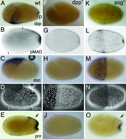
Fig. 2.
Tc-Sog transports Dpp toward the dorsal side. Lateral views of embryos at the differentiated blastoderm stage. (A–E%) Wild type (wt). (F–J%) Tc-dpp RNAi (dpp−). (K–O%) Tc-sog RNAi (sog−). (A, F, and K) Tc-dpp in situ hybridization. (B, G, and L) pMAD antibody staining. (C, H, and M) Tc-doc in situ hybridization. (D, I, and N) DAPI counterstaining of the embryos shown in C, H, and M, respectively. Serosal nuclei are bigger and wider spaced than those of the germ rudiment. (E, J, and O) Tc-pnr in situ hybridization. (A) Tc-dpp is expressed in a stripe along the border of the germ rudiment and serosa and in the primitive pit (p). (B) pMAD accumulates along the whole dorsal side of the embryo. See Results for details. Black lines indicate the germ rudiment/serosa border. (C) Tc-doc is transcribed in a subset of dorsal cells in the serosa. (D) The border of the germ rudiment and serosa is oblique (white lines indicate the dorsal and ventral point of the border). (E) Tc-pnr is expressed at the dorsal side of the germ rudiment (arrow) and in the primitive pit. (F) No Tc-dpp expression is detected. (G) pMAD could not be detected. (H) Tc-doc transcripts could not be detected. (I) The serosa/germ rudiment border is straight. (J) Tc-pnr transcripts could not be detected. (K) Tc-dpp is weakly expressed along the germ rudiment/serosa border. (L) pMAD is present in a band along the border of the germ rudiment and the serosa. Additional pMAD is found in the primitive pit. (M) Tc-doc is expressed in a DV symmetrical broad band in the serosa. (N) The germ rudiment/serosa border is straight. (O) Tc-pnr is expressed in a rim along the anterior of the germ rudiment and in the primitive pit but not at the dorsal side of the germ rudiment (arrow).

