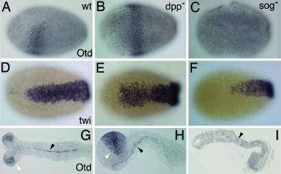
Fig. 3.
Tc-sog RNAi deletes the head; Tc-dpp RNAi enlarges the head. (A–F%) Differentiated blastoderm stages. (A–C%) Tc-Otd antibody stainings, lateral views. (D–F%) Tc-twist in situ hybridizations, differentiated blastoderm stages, ventral views. (G–I%) Tc-Otd antibody stainings; extended germ-band embryos. (A) WT. Tc-Otd is present in an anterior triangle. (B) Tc-dpp RNAi. Tc-Otd is present in a band in the anterior germ rudiment. (C) Tc-sog RNAi. Tc-Otd could not be detected. (D) WT. (E) Tc-dpp RNAi. The Tc-twist domain extends to a WT position along the AP axis but is slightly broader in the anterior half. (F) Tc-sog RNAi. The Tc-twi domain is only half as long as in the WT. (G) WT. Tc-Otd is found in the head lobes (open arrowhead) and along the ventral midline (filled arrowhead). (H) Tc-dpp RNAi, lateral view. Tc-Otd is detected along the ventral midline (filled arrowhead) and in an enlarged anterior domain (open arrowhead). (I) Tc-sog RNAi. Tc-Otd is detected only in some patches along the ventral midline (arrowhead).

