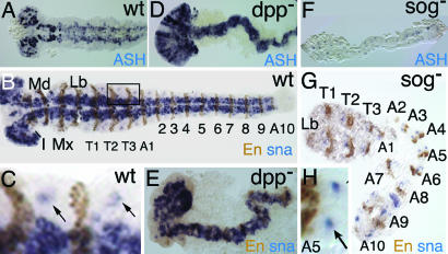
Fig. 4.
Tc-sog RNAi leads to a complete loss of the neurogenic ectoderm; extending germ-band embryos. (A–C%) WT. (D and E) Tc-dpp RNAi. (F–H%) Tc-sog RNAi. (A, D, and F) Tc-achaete-scute in situ hybridizations. (B–H%) Tc-snail in situ hybridizations (blue) with engrailed antibody staining (brown). (A) Tc-ASH is expressed in cells of the CNS and in a transverse stripe at the anterior of each segment. (B) Tc-snail is expressed in cells of the CNS. Seventeen engrailed stripes could be counted. Segments are labeled as follows: I, intercallary; Md, mandibular; Mx, maxillary; Lb, labial; T, thoracic; A, abdominal. (C) Magnification of a part of the embryo boxed in B. Tc-snail is also detected in single clusters marking the peripheral neurons of the lateral ectoderm (arrows). (D) Tc-ASH transcripts can be detected throughout the embryo. (E) Tc-snail can be detected throughout the embryo. (F) Tc-ASH can be detected only in segmental stripes. (G) Tc-snail expression is found only in single clusters. Only 13 engrailed stripes were counted. (H) Magnification of segment A5 from the embryo shown in E. The arrow points at the peripheral neurons.

