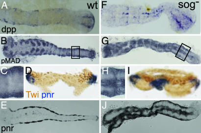
Fig. 5.
Tc-sog RNAi embryos show a double dorsal phenotype. (A–E%) WT. (F–J%) Tc-sog RNAi. (A and F) Tc-dpp in situ hybridizations. (B, C, G, and H) pMAD antibody staining. (D and I) Cross-sections with Tc-pnr in situ hybridization (blue) and Twist antibody staining marking the mesoderm (dark brown). (E and J) Tc-pnr in situ hybridizations. (A) Tc-dpp is expressed along the dorsal borders of the extending germ band and in two stripes in the growth zone. (B) pMAD is detected along the dorsal margins of the extended germ band. (C) Magnification of the area boxed in B. (D and E) Tc-pnr is expressed at the dorsal margins. (F) Tc-dpp is weakly expressed along the dorsal margins and in two strong ectopic stripes along the ventral midline. The stripes are continuous, with the stripes in the growth zone. (G) pMAD is detected along the dorsal margins of the germ band and in a strong ectopic domain along the ventral midline. (H) Magnification of the area boxed in D. (I and J) Tc-pnr is expressed along the dorsal margin and in a strong, ventral, ectopic stripe in the ectoderm.

