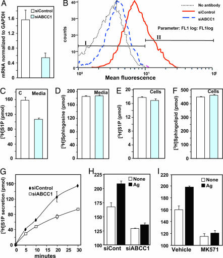
Fig. 3.
Down-regulation of ABCC1 decreases S1P secretion. RBL-2H3 cells were transfected with control siRNA or siRNA targeted to ABCC1. (A) RNA was isolated, and mRNA levels of ABCC1 and GAPDH were determined by quantitative real-time PCR. (B) In duplicate cultures, ABCC1 expression on the surface was determined by flow cytometry. Percentage of cells staining above background in fluorescence intensity gates I and II are: no antibody, 99.1 and 0.1; siControl, 73.1 and 26.8; siABCC1, 94.8 and 4.7, respectively. (C–H) RBL-2H3 cells transfected with siControl (open bars) or siABCC1 (gray bars) were labeled with [3H]Sph (1.5 μM, 0.45 μCi) for 30 min (C–F) or the indicated times (G) in serum-free medium. Lipids were differentially extracted from media (C, D, and G) and cells (E and F) into aqueous (C, E, and G) and organic fractions (D and F) and quantified by scintillation counting. Data are the means ± SD. (H) RBL-2H3 cells transfected with siControl or siABCC1 were sensitized with anti-DNP IgE overnight, labeled with [3H]Sph for 30 min, and stimulated without (open symbols) or with DNP-HSA (Ag, 100 ng/ml, filled symbols) for 30 min, and [3H]S1P released into the media was quantified. (I) RBL-2H3 cells were sensitized with anti-DNP IgE overnight, pretreated with vehicle or 15 μM MK571 for 1 h, labeled with [3H]Sph for 30 min, and stimulated without (open symbols) or with (filled bars) Ag for 30 min, and [3H]S1P released into the media was quantified. Data are the means ± SD.

