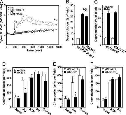
Fig. 5.
Inhibition of ABCC1 affects Ag-induced chemotaxis but not degranulation or calcium mobilization. (A) IgE-sensitized RBL-2H3 cells were loaded with the calcium indicator Fura-2AM, pretreated without or with MK571 (15 μM), and then stimulated without or with Ag (100 ng/ml), as indicated. Changes in intracellular free calcium were determined by the ratio of fluorescence emission at 510 nm after excitation at 340 and 380 nm. (B) IgE-sensitized RBL-2H3 cells were pretreated with vehicle or MK571 (15 μM) for 1 h, followed by stimulation with DNP-HSA (Ag, 100 ng/ml, filled bars) for 30 min, and degranulation was measured. (C) IgE-sensitized LAD2 cells transfected with siControl or siABCC1 were stimulated without (open bars) or with (filled bars) Ag for 30 min, and degranulation was measured. (D) IgE-sensitized RBL-2H3 cells were treated without (open bars) or with (gray bars) 15 μM MK571 for 2 h and then allowed to migrate toward vehicle (None), Ag (30 ng/ml), S1P (10 nM), fibronectin (20 μg/ml), or serum (2%) for 4 h, and chemotaxis was measured as described (13). (E and F) RBL-2H3 cells transfected with siControl (open bars) or siABCC1 (filled bars) were sensitized with anti-DNP IgE overnight and allowed to migrate toward vehicle, Ag, serum, S1P, and fibronectin for 4 h. Results are expressed as means ± SD of triplicate determinations.

