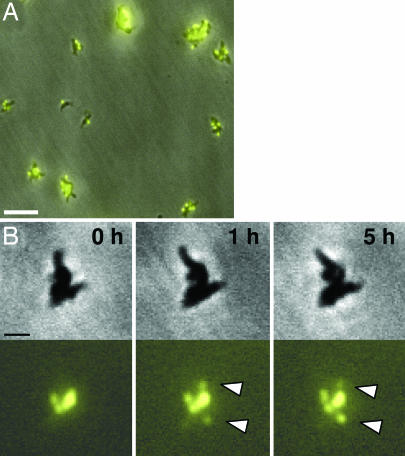
Fig. 2.
Time-lapse phase contrast/fluorescence microscopy of terminal organelle development without separation during growth of nonmotile M. pneumoniae mutant III-4. (A) Representative field for examination of nonmotile mutant III-4 plus P30-YFP. (Scale bar, 5 μm.) (B) Nonmotile mutant III-4 with selected frames over a 5-h observation during growth in a chamber slide. (Upper) Phase contrast images. (Lower) P30-YFP fluorescence images, with white arrowheads indicating new foci evident during the observation period. (Scale bar, 1 μm.)

