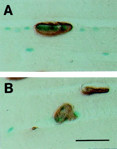
Figure 2.
Expression of AChE promoter–reporter gene constructs in synaptic compartments of mouse TA muscle fibers. (A and B) Cryostat sections stained histochemically for the simultaneous demonstration of β-gal (blue staining) and AChE (brown staining) activity. Note that the presence of blue nuclei coincides with the occurrence of neuromuscular junctions, reflecting AChE promoter activity within junctional myonuclei. (Bar = 75 μm.)

