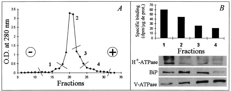
Figure 6.
Separation of M. varia microsomal fraction membranes, monitored by absorbance at 280 nm, by free-flow electrophoresis (A) and distribution of NFBS2 into the different pooled membrane fractions (B, Upper) whose composition was checked by immunodetection of protein markers (B, Lower). For the determination in B, the same amount of protein was used for each fraction. H+-ATPase, BiP, and V-ATPase are markers for PM, endomembrane, and tonoplast, respectively.

