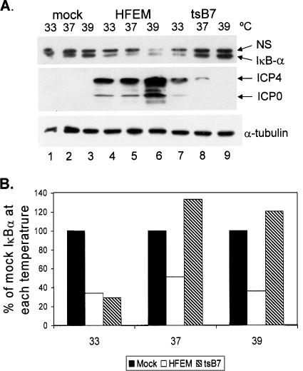
FIG. 2.
IκBα degradation is dependent on viral gene expression. (A) Replicate CV-1 cultures were mock infected or infected with HSV-1 strain HFEM or mutant tsB7 (MOI = 5) at the indicated temperatures, and lysates were prepared at 8 hpi. Western blot analysis of proteins was performed as described in Materials and Methods. Lysates fractionated on a 12% polyacrylamide gel were sequentially probed for IκBα and α-tubulin, and lysates fractionated on a 6% polyacrylamide gel were simultaneously probed for ICP0 and ICP4. Two additional independent experiments yielded similar results. NS, nonspecific. (B) Band intensities of IκBα from the Western blot in panel A were quantified using Image J as described in Materials and Methods, and the results presented are normalized to the values from mock infection. Filled bars, mock infected; open bars, HFEM infected; hatched bars, tsB7 infected.

