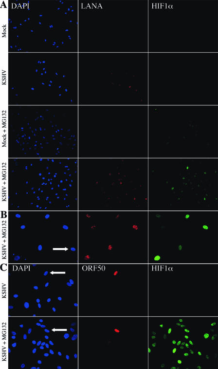
FIG. 4.
Latent KSHV-infected TIME cells stabilize more HIF1α protein than do uninfected cells. Results show the immunofluorescent detection of nucleic acid (DAPI, blue), KSHV latent nuclear antigen (LANA, red), and HIF1α (green) in mock- and KSHV-infected TIME cells 48 h postinfection in the absence or presence of the proteosome inhibitor, 20 μM MG132, for 1.5 h (magnification, ×200) (A) and 3 h (magnification, ×400) (B). The white arrow indicates an uninfected cell within the KSHV-infected population. (C) Immunofluorescent detection of nucleic acid (DAPI, blue), KSHV lytic switch protein, ORF50 or Rta (red), and HIF1α (green) in KSHV-infected TIME cells 48 h postinfection in the absence or presence of 20 μM MG132 for 2 h. The white arrows indicate the ORF50-positive cells (magnification, ×400). Panels A, B, and C are from separate experiments with similar infection rates (∼80% latent, <0.6% lytic).

