
FIG. 1.
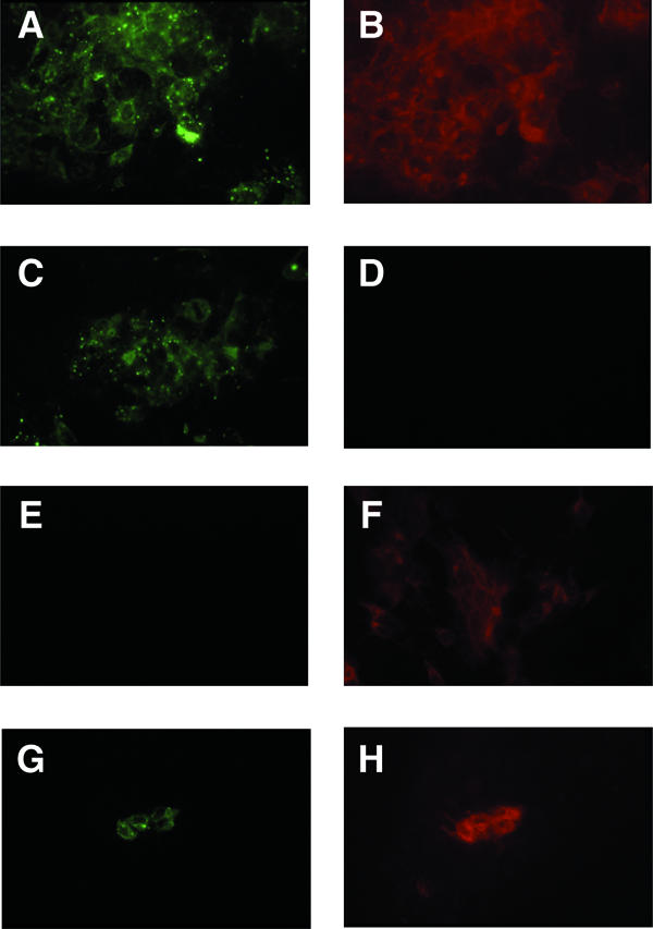
Immunofluorescence microscopy of transfected or infected 10-3 cells. Cells were stained for ORF2 protein (green) or ORF3 protein (red) after transfection (A to F) or infection (G and H). Wild-type (A, B, G, and H), ORF3-null (C and D), and ORF2-null (E and F) viruses were used. Each pair of panels represents the same field as photographed through the 25× objective, and each field contains a confluent monolayer of cells.