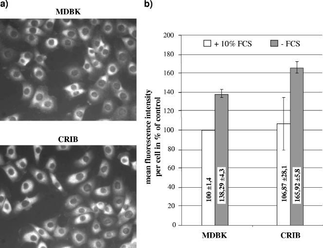
FIG. 2.
Analysis of LDL receptor expression on CRIB and MDBK cells by (a) indirect immunofluorescence assay and (b) flow cytometry. (a) CRIB and MDBK cells were grown for 16 h at 37°C and fixed with 4% paraformaldehyde in PBS. Antigen was detected by anti-LDL receptor MAb clone C7 (2.5 μg/ml) and an anti-mouse Cy3 conjugate. (b) CRIB and MDBK cells were grown either in serum-free DMEM or in DMEM containing different amounts of FCS for 16 h at 37°C, subsequently detached by being washed in PBS containing 5 mM EDTA, and fixed with 4% paraformaldehyde in PBS. Antigen was detected by anti-LDL receptor MAb clone C7 (30 μg/ml) and an anti-mouse FITC conjugate. A total number of 2,000 events were analyzed with a FACSCalibur. Data analysis was performed using the FCS Express Version 2 software (DeNovo Software). Electronic gates were set according to the negative control included in each test series, defining less than 2% of the cells as positive.

