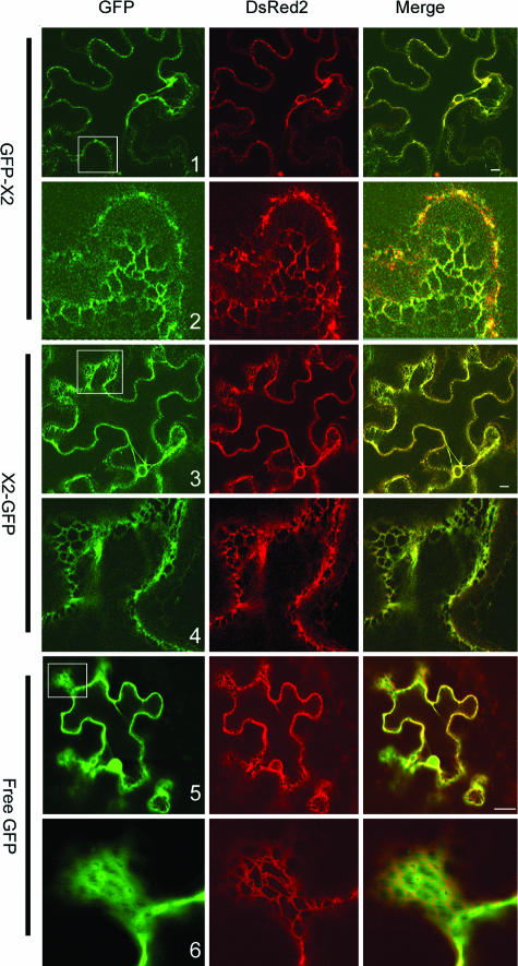
FIG. 4.
Subcellular localization of GFP-X2 and X2-GFP. GFP fusions and ER-dsRed2 (an ER marker) were expressed in leaves of N. benthamiana by using agroinfiltration as described in Materials and Methods. Epidermal cells were examined 3 days after agroinfiltration by confocal microscopy. In the merge panel, the colocalization of the GFP fluorescence (green) and of the ER marker fluorescence (red) results in a yellow color. Panels 2, 4, and 6 are close-up views of regions included in the white squares in panels 1, 3, and 5. Bars on the merged images represent 10 μm.

