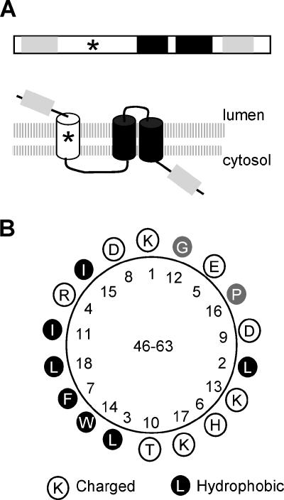
FIG. 8.
Topological model of X2 in ER membranes. (A) Proposed topological model of X2. On the top of the panel is the linear representation of membrane association domains of X2. The light gray regions represent hydrophobic domains (TM1 and TM4, as in Fig. 1) that do not traverse the membranes. Transmembrane α-helices TM2 and TM3 are shown by the black boxes as in Fig. 1. The star represents a putative amphipathic helix. Below the domain diagram is the topological model of X2 in ER membranes. The double-lipid layer of the membranes is represented by the two shaded horizontal lines. The predicted orientations of the various transmembrane domains within the membrane are shown. (B) Helical wheel projection of a putative amphipathic helix located between amino acids 46 and 63.

