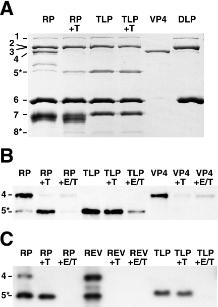
FIG. 6.
Trypsin cleavage of free and assembled VP4. In each panel, protein bands are identified by viral protein number on the left. (A) Trypsin cleavage of recoated and authentic particles. A Coomassie-stained SDS-PAGE gel is shown. “RP” indicates purified recoated particles from pool d in Fig. 5. “+T” indicates analytical trypsin digestion (see Materials and Methods). The TLPs were prepared with trypsin in the cell culture medium. Samples of purified recombinant VP4 and DLPs are shown for comparison. (B) Effect of assembly and uncoating on the trypsin cleavage of VP4. A Western blot with monoclonal antibody HS2, which recognizes VP5* and VP4, is shown. Samples are labeled as in panel A. “+E/T” indicates that EDTA was added to 5 mM before trypsin digestion. The less-efficient degradation of VP4 in the presence of EDTA (last lane) reflects mild inhibition of trypsin by EDTA. (C) Effect of order of addition during recoating on trypsin sensitivity of VP4. A Western blot with monoclonal antibody HS2 is shown. “RP” indicates a standard recoating reaction mixture, in which VP4 was added before VP7. “REV” indicates a reverse order of addition, in which VP7 was added before VP4. In this panel, complete recoating mixtures, which also include unassembled VP4 and VP7, were analyzed.

