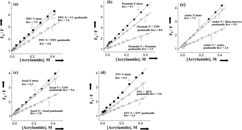
FIG. 5.
Stern-Volmer analysis of N-associated tryptophan quenching. Plots depicting tryptophan fluorescence quenching of trimeric N protein in the presence of neutral quencher acrylamide are shown. A solution containing 25 nM trimeric N was treated with an excitation wavelength of 292 nm, and emission was recorded at 330 nm. The fluorescence intensity at 330 nm for Prospect Hill virus (PHV; a), Puumala virus (b), Seoul virus (c), SNV (d), and Andes virus (e) was determined by using different input concentrations of acrylamide. Plots of F0/F as a function of acrylamide concentration (Stern-Volmer plots) for free N protein trimer in the absence of any panhandle (▪) and in the presence of minipanhandle from the same (○) or a different virus (□). The panhandle RNA concentration was three times the dissociation constant for the panhandles used in the experiment. The Stern-Volmer plots of N protein trimers in the presence of different panhandles is shown by arrows, and the corresponding Ksv values are also presented. The same method was used to calculate the Ksv values for the interaction of other N protein trimers with different panhandles. These data are presented in Table 4. TSW, tomato spotted wilt virus; RVF, Rift Valley fever virus.

