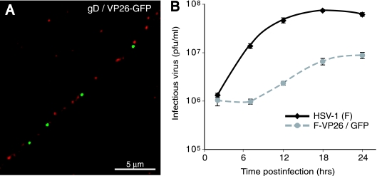
FIG. 4.
Localization of capsids and glycoproteins in human SK-N-SH neurons infected with HSV-1 F-VP26-GFP. (A) SK-N-SH neurons were infected with F-VP26-GFP for 24 h, fixed, permeabilized, and stained with rabbit polyclonal anti-gD antibodies followed by Texas red-conjugated donkey anti-rabbit IgG. Capsids are depicted in green, and gD is depicted in red. Scale bar, 5 μm. (B) Growth of F-VP26/GFP on differentiated SK-N-SH cells. SK-N-SH neurons were infected with either VP26/GFP or wild-type HSV strain F using 5 PFU/cell, and the combined cells and cell culture supernatants were collected at 2, 8, 12, 18, and 24 h. The titer of infectious virus was determined on Vero cells.

