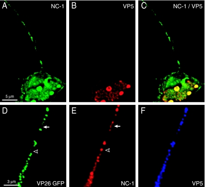
FIG. 9.
Anti-NC-1 antibodies stain HSV proteins other than those in capsids. (A to C) SK-N-SH neurons were infected with HSV TsProt.A for 18 h, fixed, permeabilized, and stained with a combination of pooled mouse anti-VP5 MAb (red) and preabsorbed rabbit polyclonal anti-NC-1 antibodies (green) followed by Texas red-conjugated donkey anti-mouse IgG and FITC-conjugated donkey anti-rabbit IgG secondary antibodies. (D to F) SK-N-SH neurons infected with F-VP26/GFP for 24 h were fixed, permeabilized, and simultaneously stained with preabsorbed rabbit polyclonal anti-NC-1 antibodies and anti-VP5 mouse MAb and then with secondary antibodies: Texas red-conjugated donkey anti-mouse IgG and Cy5-conjugated donkey anti-rabbit IgG. VP26/GFP-labeled axons are shown in green, NC-1 is shown in red (pseudoimaged from Cy5 blue), and VP5 is shown in blue (pseudoimaged from Texas red). The arrow and arrowhead indicate examples of NC-1 staining which did not overlap with the VP26/GFP signal (green).

