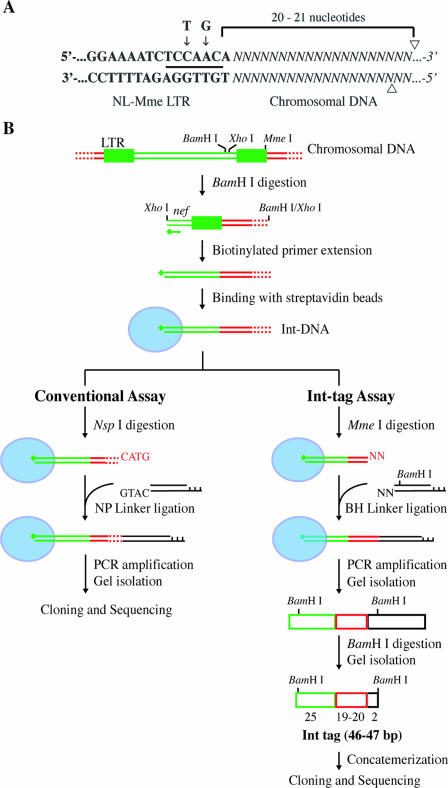
FIG. 1.
Assays for genome-wide analysis of HIV-1 integration sites. (A) Construction of mutant HIV with a type IIS MmeI restriction site in the LTR. Bold letters denote viral DNA sequences at the U5 region of the LTR, and italicized letters denote chromosomal DNA. The nucleotides at the positions indicated by the arrows were changed from T and G in the wild-type sequence to C and A, respectively, in the NL-Mme mutant, generating a new recognition site for MmeI (underlined). Arrowheads indicate cleavage sites for MmeI. (B) Schematic diagram outlining the major steps of the conventional assay and the high-throughput Int-tag assay. Viral, cellular, and linker DNAs are denoted by green, red, and black lines or boxes, respectively. Red dotted lines denote cellular DNA with various lengths. Blue ovals represent streptavidin beads, and green diamonds represent biotin. See Materials and Methods for a detailed description of the experimental procedures.

