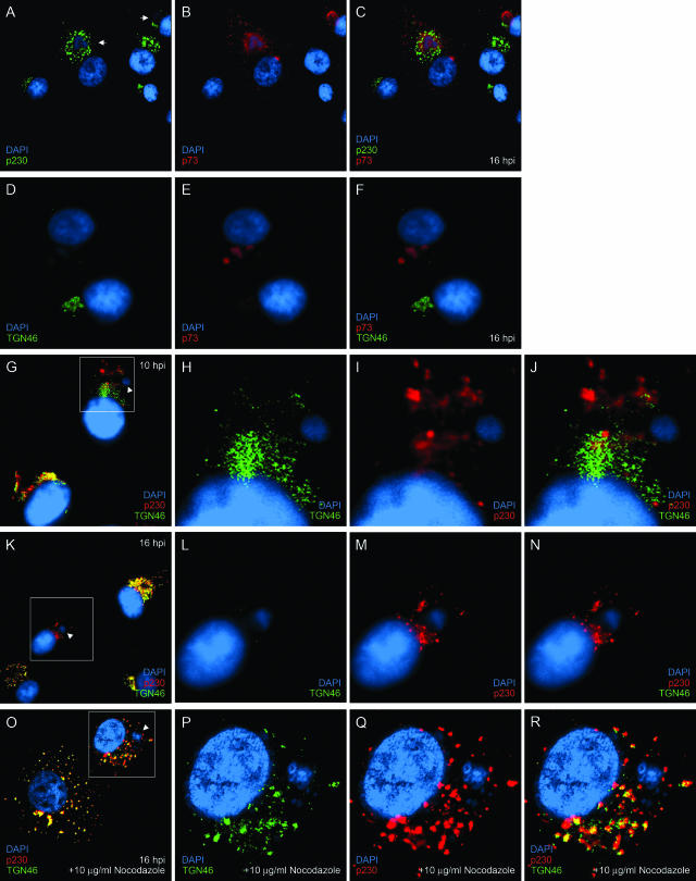
FIG. 1.
The effect of ASFV on TGN is microtubule dependent. Vero cells grown on coverslips were infected for 16 h with the Vero cell-adapted Badajoz 1971 (Ba71v) strain of ASFV (A to N) or infected for 8 h with Ba71v and incubated for a further 8 h in the presence of 10 μg/ml nocodazole (O to R) and then fixed, permeabilized, and processed for indirect immunofluorescence. The cells were stained with p230 antiserum (A) or sheep anti-TGN46 (D, G, K, and O) and 4H3 (B and E) or p230 antiserum (G, K, and O) and then with appropriate secondary antibodies conjugated to Alexa-488 or -594. All cells were incubated with DAPI (4′,6′-diamidino-2-phenylindole) dye. The cells were viewed at 60× magnification (1.4 normal aperture) with a Nikon E800 microscope. Images were captured with a Hamamatsu C-4746A charge-coupled-device camera and were deconvolved and digitally merged with Improvision Openlab 2.1.3 software. Twenty optical sections 0.2 μm thick were analyzed. The images were resized and annotated using Adobe Photoshop CS 8. Arrows indicate recruitment of p230 to viral factories, arrowheads indicate viral factories, and boxed areas designate the region enhanced in the subsequent threepanels.

