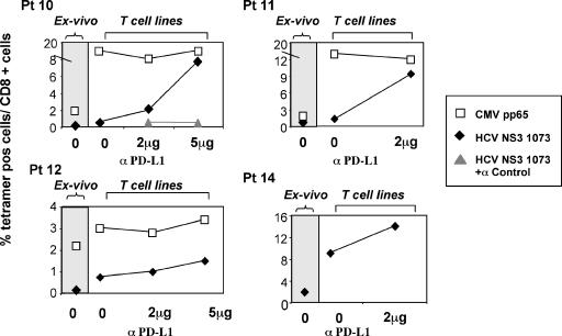
FIG. 4.
Effect of anti-PDL-1 antibody on peptide-induced proliferation of HCV-specific CD8 cells. Frequencies of tetramer-positive (tetramer-pos) CD8 cells are illustrated ex vivo (gray area) and upon specific peptide stimulation of T-cell lines (white area) in the absence or in the presence of different anti-PDL-1 concentrations. For these experiments, adherent mononuclear cells isolated from peripheral blood mononuclear cells of patients with chronically evolving infection were incubated for 45 min with different concentrations (2 or 5 μg/ml) of anti-PD-L1 (e-Bioscience, Boston, MA) or control antibody (negative control for mouse immunoglobulin G1 Ab-1; Fremont, CA). Cells were then washed and coincubated for 10 days with nonadherent cells and specific peptides (HCV NS3 1073-1081 or CMV pp65) at the final concentration of 1 μg/ml (interleukin 2 was added on day 3). White squares (□) indicate tetramer-positive cells specific for CMV pp65 (NLVPMVATV); black rhombs (⧫) indicate tetramer-positive cells specific for HCV (CINGVCWTV); gray triangles ( ) indicate HCV NS3 1073 tetramer-positive CD8 cells stimulated with the specific peptide in the presence of a control isotype antibody.

