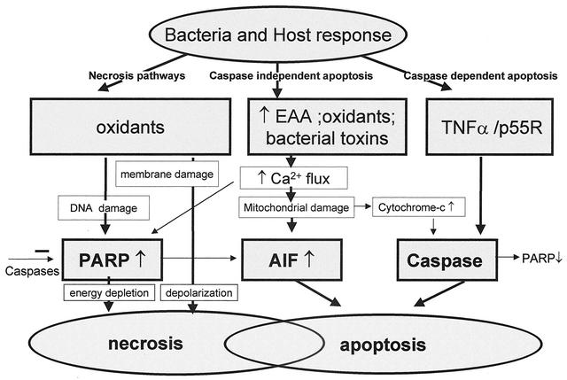
FIG. 5.
Neuronal cell death pathways may be divided into necrotic pathways, caspase-independent apoptotic pathway, and caspase-dependent apoptotic pathway. Bacterial and host oxidants cause damage to cell membranes via lipid peroxidation, leading to loss of membrane integrity and depolarization and finally necrotic cell death. Oxidants also cause DNA damage, resulting in the energy-consuming activation of the poly(ADP-ribose) polymerase (PARP). When DNA repair is futile because of the magnitude of the damage, massive energy depletion will cause necrotic cell death. Oxidants, high concentrations of excitatory amino acids (EAA), and bacterial toxins such as the pneumolysin of S. pneumoniae or the hemolysin of S. agalactiae all cause increased cytosolic free calcium levels. This may result in PARP activation and contribute to necrotic death but primarily causes damage to the mitochondrial outermembranes with PARP-dependent release of apoptosis-inducing factor (AIF). Free cytosolic apoptosis-inducing factor will move into the nucleus and cause chromatin condensation and apoptotic cell death. Release of inflammatory mediators such as TNF-α in response to the invading bacterial pathogens will result in activation of caspases. This results in activation of the apoptotic pathway and will also inhibit necrosis through inactivation of PARP. Release of cytochrome c from mitochondria following mitochondrial damage in response to increased cytosolic free calcium levels also causes activation of caspases and apoptotic death. As indicated in the figure, several crossroads exist between the different death pathways. In many cases, both necrotic and apoptotic cell death may be revealed on histologic examination.

