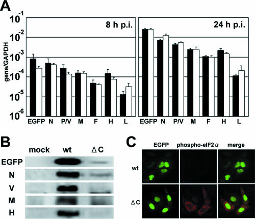
FIG. 2.
Translational inhibition in MVΔC-infected cells. (A) Quantification of viral transcripts in a single step of growth. A549/hSLAM cells were infected with MVwt or MVΔC at an MOI of 0.5 and incubated in the presence of a fusion-blocking peptide. At 8 and 24 h p.i., the EGFP, N, P/V, M, F, H, and L mRNAs in the virus-infected cells were quantified by RT-QPCR. All data were normalized by the corresponding level of GAPDH mRNA. Filled bars, MVwt; open bars, MVΔC. The data represent the means ± standard deviations of triplicate samples. (B) Detection of viral proteins by immunoblotting. A549/hSLAM cells were infected with MVwt or MVΔC at an MOI of 0.5 and incubated in the presence of a fusion-blocking peptide. At 24 h p.i., EGFP and the N, V, M, and H proteins in the virus-infected cells were detected by immunoblotting using appropriate primary and secondary antibodies. (C) Immunofluorescence staining of phospho-eIF2α in MV-infected cells. A549/hSLAM cells were infected with MVwt or MVΔC at an MOI of 0.5 and incubated in the presence of a fusion-blocking peptide. At 36 h p.i., the cells were fixed and permeabilized, and phospho-eIF2α was detected by indirect immunofluorescence staining using a phospho-eIF2α-specific antibody. Green and red fluorescence indicates EGFP and phospho-eIF2α, respectively.

