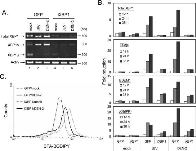
FIG. 6.
siRNA targeting of XBP1 mRNA attenuated virus-induced ER expansion and XBP1 downstream gene induction. (A) Stable N18 cell populations transduced with lentivirus expressing GFP or iXBP1-GFP were mock infected or infected with JEV or DEN-2 (MOI, 5) for 24 or 30 h, respectively, before XBP1 mRNA expression and splicing were analyzed by RT-PCR and PstI digestion, as described in the legend to Fig. 1. (B) Cells were harvested at 12, 24, and 36 h post-virus infection (MOI, 5) for real-time RT-PCR analysis using primer sets specific for the indicated genes, as described in the legend to Fig. 1. (C) N18 cells transduced with lentivirus encoding GFP or iXBP1-GFP were mock infected or infected with DEN-2 (MOI, 5) for 30 h. Flow cytometry analysis of the ER contents was then performed with brefeldin A and BODIPY 558/568 conjugate staining, followed by analysis using FACSort (Becton Dickinson) and CellQuest software.

