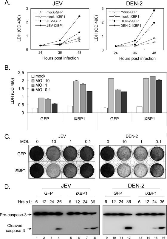
FIG. 8.
Functional impairment of XBP1 in N18 cells exacerbates CPE induced by JEV and DEN-2. (A) N18 cells stably transduced with GFP or iXBP1-GFP lentivirus were mock infected or infected with JEV or DEN-2 at an MOI of 1. The culture supernatants were collected for LDH release assay at the indicated time points. The data shown are means ± standard deviations from three independent experiments. OD 490, optical density at 490 nm. (B and C) The cells described for panel A were mock infected or infected with JEV or DEN-2 at various doses (MOIs as indicated) for 48 h. The culture supernatants were collected for LDH release assay (B), and the surviving cells were fixed and stained with crystal violet (C). The LDH readings are the means plus standard deviations from two independent experiments. (D) Cells were infected with JEV or DEN-2 (MOI, 5) and harvested at the indicated time points for caspase 3 activation analysis by immunoblotting. p.i., postinfection.

