FIG. 4.
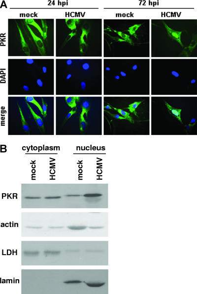
PKR accumulates in the nucleus during HCMV infection. (A) HFs were mock infected or infected with HCMV (MOI = 3). At the indicated times postinfection, indirect immunofluorescence for endogenous PKR was performed using a PKR mouse monoclonal antibody, and PKR was visualized using fluorescence microscopy. (B) HFs were mock infected or infected with HCMV. At 72 hpi, cell lysates were fractionated into cytoplasmic and nuclear components, and immunoblot analyses of PKR (PKR B-10 mouse monoclonal antibody), actin, lactate dehydrogenase (LDH), and lamin B were performed.

