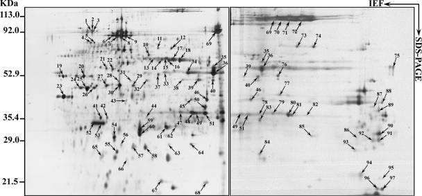
FIG. 2.
2D gel electrophoresis patterns of proteins. Acanthamoeba polyphaga mimivirus was separated using 18-cm pI 4 to 7 (left) or 6 to 11 (right) IPG strips for the first dimension (IEF) followed by 10% linear SDS-PAGE for the second dimension. Spots revealed by silver staining were cut out from the gel and subjected to trypsin digestion followed by MALDI-TOF-MS analysis. Molecular sizes and pI ranges are indicated. Identified proteins are listed in Table 1 (also see details in Table S1 in the supplemental material).

