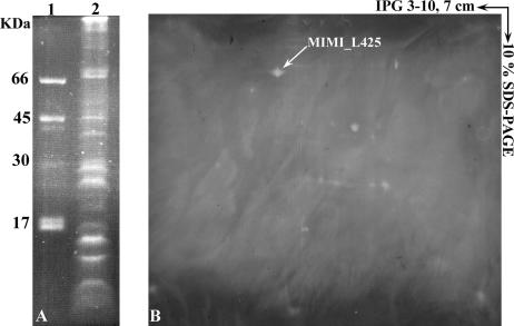
FIG. 3.
Glycosylation pattern of Acanthamoeba polyphaga mimivirus proteins detected by fluorescence assay. Mimivirus proteins were separated by 10% SDS-PAGE (A) or by 2D electrophoresis using 7-cm IPG strips (pI 3 to 10) (B), and glycosylated proteins were revealed with GlycoProfile III. Lane 1, standard glycosylation markers; lane 2, mimivirus.

