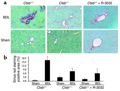
Figure 5.
Hepatic fibrosis is increased in Ctsb+/+ BDL compared with Ctsb–/– BDL mice. (a) Two weeks after the surgical procedure, liver tissue was obtained from BDL and sham-operated Ctsb+/+, Ctsb–/–, and R-3032–treated Ctsb+/+ BDL mice, and collagen fibers were stained with sirius red as described in Methods. (b) The surface area stained with sirius red was quantitated using digital image analysis. Sirius red staining was quantitatively greater in Ctsb+/+ BDL than in Ctsb–/– and R-3032–treated Ctsb+/+ BDL mice (P < 0.001, n = 4 for each group). Only minimal sirius red staining was observed in sham-operated mice from the three groups of animals. (Original magnification ×20.)

