Figure 2.

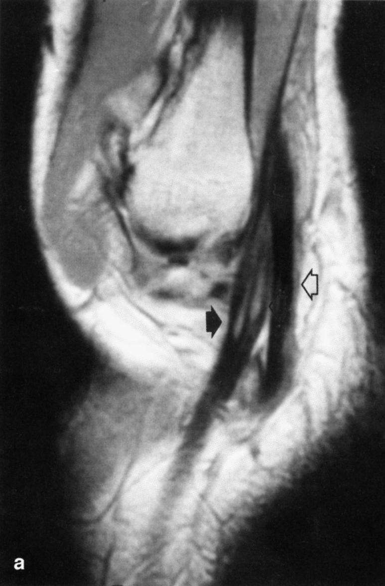
Sagittal oblique spin-echo proton density-weighted magnetic resonance image showing high signal intensity within the peroneus brevis tendon at the lateral malleolar level (black arrow). The peroneus longus tendon (white arrow) is normal.

Sagittal oblique spin-echo proton density-weighted magnetic resonance image showing high signal intensity within the peroneus brevis tendon at the lateral malleolar level (black arrow). The peroneus longus tendon (white arrow) is normal.