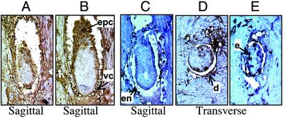
Fig. 4.
Immunological determination of FAS expression in developing embryos with anti-FAS antibodies. Timed matings were performed and the uteri were resected at E6.5 (A and B) and E5.5 (C–E) postcoitus. The uteri were fixed in Bouin's solution and sectioned through the decidua-containing regions. The sections were treated with anti-FAS goat antibodies, and the immune complexes were detected immunohistochemically with peroxidase-conjugated secondary antibodies (dark brown regions). (A) Small, growth-retarded E6.5 embryo. (B) Normal-looking E6.5 embryo. (C and D) Sagittal and transverse sections of normal-looking E5.5 embryos. (E) Growth-retarded E5.5 embryo. (Magnification: ×400.) Arrows indicate some of the regions expressing high FAS levels. epc, ectoplacental cone; vc, visceral endoderm; en, endoderm; d, decidual cells; e, ectoderm.

