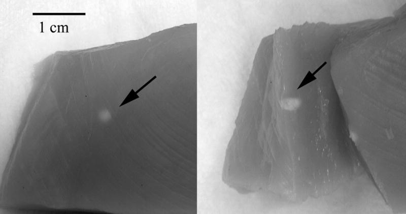
Figure 4.

Photograph of lesion site in chick breast. In this case, the lesion was produced from a 15 second exposure at 5600 W/cm2. The external view (left) shows a typical blanched spot at the lesion site. The cross sectional view (right) shows tissue alteration to a depth of several millimeters.
