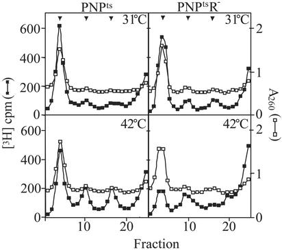
Fig. 4.
Sedimentation analysis of ribosomes. Cultures were grown as described in Fig. 1 except that 5 μCi/ml [3H]uridine was added to the fresh YT medium at 42°C. 3H-labeled S30 extracts (≈20,000 cpm) were layered on 11-ml 5–20% sucrose gradients and centrifuged at 18,500 rpm in an SW41 rotor (Beckman) for 15 h at 4°C. Fractions were collected from the bottom, and 3H counts were determined by scintillation counting. An excess of unlabeled, wild-type extract was present in each gradient to determine the positions of the 70S, 50S, and 30S peaks (shown by arrows) by absorption at 260 nm. Total cpm in all of the samples were normalized to the same value to correct for loading and recovery variations.

