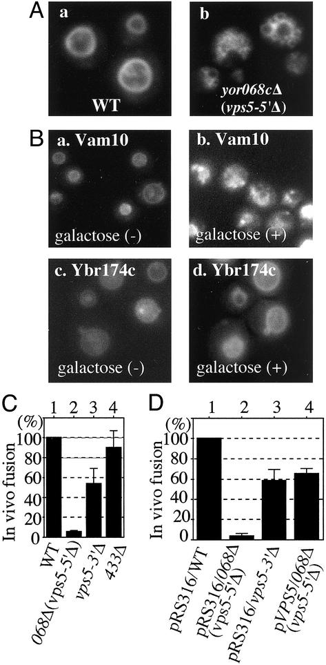
Fig. 1.
Vacuole fusion in vivo. (A) Wild-type (WT) strain BY4742 (a) and yor068cΔ (b) were grown in 0.2 ml of yeast extract/peptone/dextrose (YPD) in 15-ml culture tubes at 30°C. After 16 h, 0.4 ml of YPD with 5 μM FM4–64 (15) was added, incubation was continued for 4 h at 30°C, and cells were photographed (7). (B) Vacuole morphology before and after induction of Vam10p (a and b) and Ybr174p (c and d) by galactose. (C) Osmotic stress-induced vacuole fusion. WT strain BY4742 (lane 1) and yor068Δ (lane 2), vps5-3′Δ (lane 3), and ydr433wΔ strains were grown in 0.5 ml of YPD in 15-ml culture tubes at 30°C. After 16 h, 1 ml of YPD with 5 μM FM4–64 was added and incubation was continued at 30°C until OD600 = 5 (4–5 h). Samples (400 μl) were centrifuged (6,000 × g for 30 sec) and cells were resuspended in 350 μl of water. After 3 min at 23°C, in vivo vacuole fusion events were counted for 4 min in eight fields (30 sec per field). The average and SD of five experiments are presented. (D) Vector pRS316-transformed WT strain BY4742 (lane 1) and yor068cΔ (lane 2), vps5Δ (lane 3), and pVPS5-transformed yor068cΔ (lane 4) strains were grown in 0.5 ml of SD-ura medium and analyzed as above.

