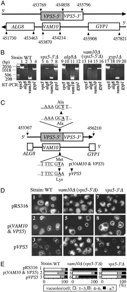
Fig. 3.
Relationship of VPS5 and VAM10. (A) Map of the ALG8, VAM10, VPS5, GYP1 region. The VPS5 gene encompasses 2,028 bp (shaded plus open boxes in the figure), from 453769 to 455796 (19, 20), although only residues 454858–455796 were removed in the vps5Δ strain. The VAM10 gene lies within the VPS5 gene on the opposite strand. (B) Levels of mRNA from ALG8, VAM10, VPS5, and GYP1 genes in BY4742 and BY4742 mutants bearing deletions, which were assayed by RT-PCR by using the oligonucleotides listed in C for VPS5 and VAM10 and GGCTCCATTGTCGCCG (5′-ALG8), TCACTGCCATAGTAATTCGT (3′-ALG8), TACACAGGCCTGTTGTTTG (5′-GYP1), and TTACAGCCAGTGCGACGT (3′-GYP1). RNA preparation used RNase-free DNase I and TRIzol (Invitrogen). Samples also were analyzed without the RT step to confirm the RNA origins of the RT-PCR products (data not shown). (C) Plasmids for gene expression were prepared as described in Methods. (D) Vacuole morphology was examined in WT, vam10Δ (vps5-5′Δ), and vps5-3′Δ strains bearing the control plasmid (pRS316), p(VAM10 and VPS5), or pVPS5 prepared as in B above, which were grown in 0.5 ml of SD-ura medium in 15-ml tubes at 30°C. After 16 h, 1 ml of YPD with 5 μM FM4–64 was added, incubation was continued for 4 h, and vacuole morphology was examined. Pictures of multiple fields were taken for each condition. (E) For each of the strains shown in D, the percentage that have one to three, four to six, or more than seven vacuoles was scored for 120–160 cells.

