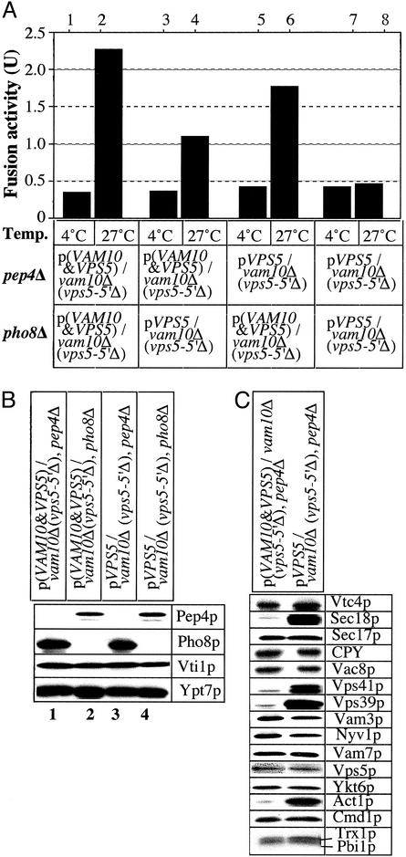
Fig. 4.
Fusion activity and protein composition of vacuoles from vam10Δ cells. (A) Standard fusion assays were performed with vacuoles from VAM10, pep4Δ; VAM10, pho8-Δ; vam10-Δ, pep4-Δ; and vam10-Δ, pho8-Δ strains. (B and C) Vacuoles (6 μg) from each indicated strain were analyzed by immunoblotting.

