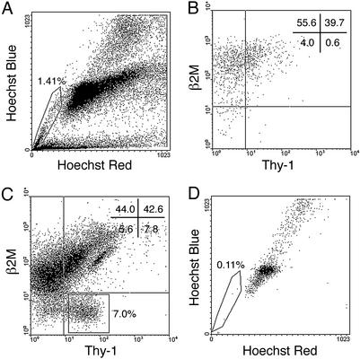
Fig. 4.
Flow cytometric analysis of cryptorchid testis cells stained with Hoechst 33342 followed by antibody staining for β2M and Thy-1. (A) Hoechst staining pattern of cryptorchid testis cells. Testis SP cells are enclosed in box. The testis SP disappeared in the presence of verapamil (data not shown). (B) β2M vs. Thy-1 expression profile of the testis SP gated in A, with quadrant statistics. There are few β2M–Thy-1+ cells in the testis SP cell fraction. (C) Staining profile of Thy-1 vs. β2M for cryptorchid testis cells. β2M–Thy-1+ cells (≈7%) are gated in the rectangle. (D) Hoechst emission profile of β2M–Thy-1+ cells gated in C is shown. SP cells and β2M–Thy-1+SP– cells were sorted simultaneously for transplantation assay. The number of spermatogenic colonies generated by 105 cells transplanted to recipients was: SP in A = 0, n = 18; β2M–Thy-1+ in C = 76 ± 15; unsorted cells = 4.7 ± 0.7, n = 18 (mean ± SEM).

