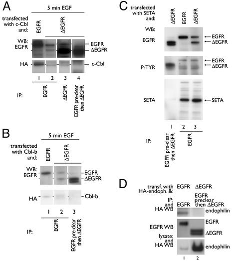
Fig. 2.
ΔEGFR does not interact with c-Cbl, Cbl-b, SETA, or endophilin. (A) HEK293 cells were transfected with c-Cbl and EGFR (lane 1) or ΔEGFR (lanes 2–4), serum-starved overnight, and incubated with 100 ng/ml EGF for 5 min. EGFR IP (Ab-1, Oncogene Science) revealed one band corresponding to wild-type EGFR (lane 1), or two bands corresponding to EGFR and ΔEGFR when cells were transfected with ΔEGFR (lane 2). When a mAb 806 IP was prepared from cells transfected with ΔEGFR, two bands were obtained, with the higher, less-intense band corresponding to wild-type EGFR (lane 3). When anti-EGFR preclearing preceded mAb806 IP, only one band corresponding to ΔEGFR was obtained (lane 4). c-Cbl was found in IPs when EGFR, but not ΔEGFR, was present (lanes 1–4). (B) Similar experiments with HEK293 cells transfected with Cbl-b showed Cbl-b in wild-type but not ΔEGFR-specific IPs (lanes 1–3). (C) Similar studies in cells grown in the continuous presence of serum demonstrated that SETA was found in IPs from HEK293 cells that contained wild-type EGFR (lanes 2 and 3), but not ΔEGFR alone (lane 1). A phosphotyrosine blot verified that receptors were activated. Please note that IPs in lanes 1 and 3 were from the same lysates, and so SETA protein was expressed in the sample from which lane 1 was prepared. (D) Although expression levels of endophilin A1 were higher when it was cotransfected with ΔEGFR than with EGFR (compare lanes 1 and 2), it was not present in ΔEGFR-specific IP (lane 2), but was present in EGFR IP (lane 1) as expected. Differences in endophilin A1 levels may relate to its higher turnover when EGFR is cotransfected.

