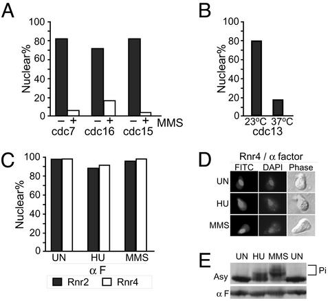
Fig. 6.
Cell cycle effect on Rnr2 and Rnr4 redistribution induced by genotoxic stress. (A) Change of Rnr4 nuclear localization in cdc mutants treated with MMS. cdc7-1, cdc16-123, and cdc15-2 cells were arrested at 37°C for 2 h, treated with 0.02% MMS for an additional 2 h, and processed for IMF. The percentage of cells displaying a predominantly nuclear Rnr4 signal was calculated by counting 200 cells per experiment, each done in duplicate. (B) Loss of Rnr4 nuclear localization induced by cdc13-1-mediated DNA damage. A cdc13-1 culture was grown at 23°C to early log phase and shifted to 37°C for 2 h. Cells were quantified as described in A. (C) No change in Rnr2 and Rnr4 localization in G1 cells treated with HU or MMS. Asynchronous cultures were blocked in G1 with α-factor (α F). The arrest was maintained while cells were treated with 150 mM HU or 0.02% MMS for 2 h, and then processed for IMF. The percentages of each cell population with a predominantly nuclear signal for Rnr2 (black bar) or Rnr4 (white bar) were quantified as described in A.(D) A representative collage of Rnr4 IMF images in MMS-treated, α-factor-arrested G1 cells. (E) Comparison of HU- and MMS-induced Rad53 phosphorylation between cells in an asynchronous (Asy) culture and G1-arrested (α F) cells.

